Home
Uncategories
Hip And Leg Bone Diagram - Pelvis Definition Anatomy Diagram Facts Britannica / Each circuit displays a distinctive voltage condition.
Hip And Leg Bone Diagram - Pelvis Definition Anatomy Diagram Facts Britannica / Each circuit displays a distinctive voltage condition.
Hip And Leg Bone Diagram - Pelvis Definition Anatomy Diagram Facts Britannica / Each circuit displays a distinctive voltage condition.. Its lower end helps create the knee joint. When you stand or walk, all the weight of your upper body rests on them. Hip and thigh bones joints muscles kenhub. The knee joint is the largest joint in the body and is primarily a hinge joint, although some sliding and rotation occur. Femur bone diagram, picture of femur bone diagram.
The ball and socket bony structure. The femur, or thighbone, is the longest and largest bone in the human body. Muscles, tendons, and ligaments run along the surfaces of the feet, allowing the complex movements needed for motion and balance. Posted on april 18, 2019april 18, 2019. The hip itself is a ball and socket joint, much like the shoulder.
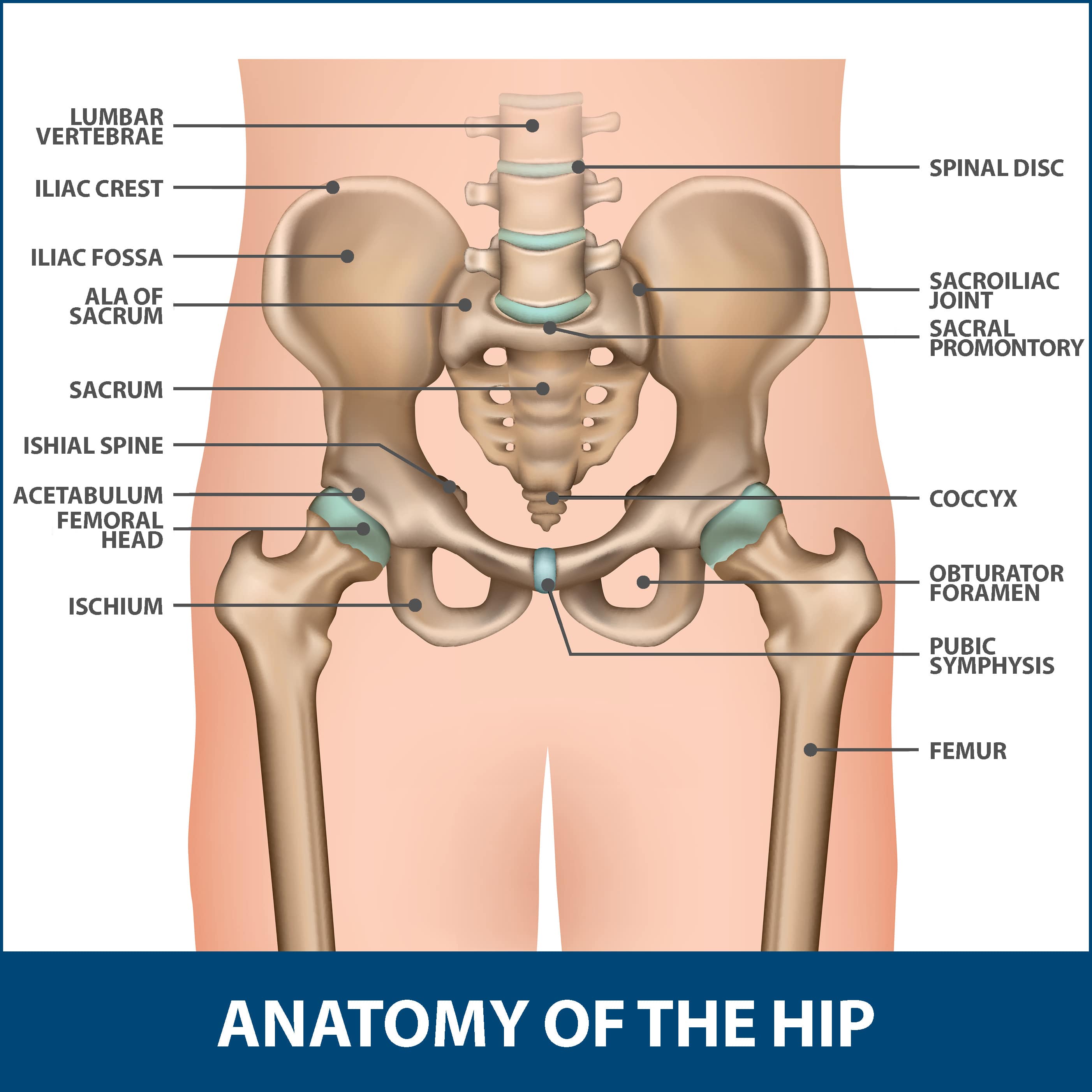
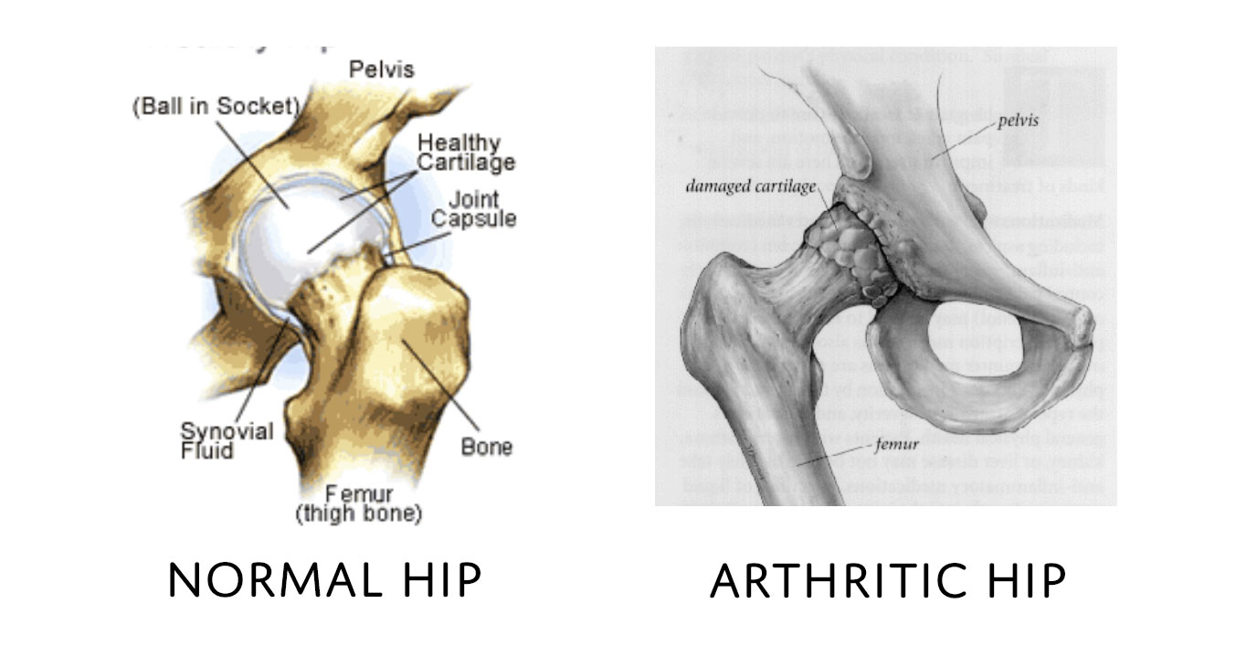
Hip Fractures Information Florida Orthopaedic Institute from www.floridaortho.com Diagram b shows that abdominal support actually lifts the front of the pelvis into proper vertical motions of the hip under the trunk. The femur is the upper leg bone or thigh. The hip bone (os coxae, innominate bone, pelvic bone or coxal bone) is a large irregular bone, constricted in the center and expanded above and below. Part of the reason for the hips stability is that there is a very deep socket called the acetabulum in the hip joint. The medial muscles of the hip are involved in the adduction of the leg i.e. The calcaneus (heel bone) is the largest bone in the foot. These bones fit uniquely with other, especially, the way they lock and unlock themselves when the foot moves from one direction to the other. Bringing the leg back towards the midline.
Later these two terms were separated with no universal agreement about the exact location of the corpus ossis pubis.
The hip bone (os coxae, innominate bone, pelvic bone or coxal bone) is a large irregular bone, constricted in the center and expanded above and below. Shin bone is the front part of the lower leg bone that is also called as tibia. The bone surfaces of the femoral head and acetabulum have a smooth durable layer of articular cartilage that cushions the ends of the bones and allows for smooth movement. Ditulis oleh anonim rabu, 07 agustus 2019 tambah komentar edit. Hip muscle strains info florida orthopaedic institute. Part of the reason for the hips stability is that there is a very deep socket called the acetabulum in the hip joint. 2006 kia optima belt diagram. Want to learn more about it? The medial muscles of the hip are involved in the adduction of the leg i.e. The piriformis muscle is what lets the hip rotate laterally, which is necessary in order for the legs to cross. The femur is the upper leg bone or thigh. Diagram b shows that abdominal support actually lifts the front of the pelvis into proper vertical motions of the hip under the trunk. Your leg bones are the longest and strongest bones in your body.
Muscles of hip, thigh, leg, and foot. Admin maret 17, 2021 another form of diagram is called the domain diagram, which is a diagram that illustrates the relationship between different. The femur, or thighbone, is the longest and largest bone in the human body. When you stand or walk, all the weight of your upper body rests on them. The ball and socket bony structure.
Total Hip Replacement Dr Peter Walker from drpeterwalker.com.au The wiring diagram on the opposite hand is particularly beneficial to an outside electrician. There are numerous structures that contribute stability to the hip: When you stand or walk, all the weight of your upper body rests on them. 2006 kia optima belt diagram. These muscles include the adductors (adductor magnus. Learn about hip and leg bones with free interactive flashcards. The hip bone (os coxae, innominate bone, pelvic bone or coxal bone) is a large irregular bone, constricted in the center and expanded above and below. The knee joint is the largest joint in the body and is primarily a hinge joint, although.
Each circuit displays a distinctive voltage condition.
Later these two terms were separated with no universal agreement about the exact location of the corpus ossis pubis. The piriformis muscle is what lets the hip rotate laterally, which is necessary in order for the legs to cross. Tensor fascia lata trigger point in it band and hip pain dr perry details the tensor fascia late trigger point that cause hip pain and it band syndrome hip injuries hip disorders take a look at some mon and not so mon hip injuries and. The foot bones shown in this diagram are the talus, navicular, cuneiform, cuboid, metatarsals and calcaneus. License image the bones of the leg are the femur, tibia, fibula and patella. Its lower end helps create the knee joint. Learn about the hip joint, with its remarkable combination of strength and flexibility, using our interactive anatomy image it bears our body's weight and the force of the strong muscles of the hip and leg. The bones of the leg are the femur, tibia, fibula and patella. Cited after worker's leg amputated. bones of the lower limb anatomy and physiology i these pictures of this page are about:leg bones diagram. The bone surfaces of the femoral head and acetabulum have a smooth durable layer of articular cartilage that cushions the ends of the bones and allows for smooth movement. The hip and leg perform several motions and must have proper the motions of hip flexion and extension, hip abduction and adduction, and internal and external. The tarsal bones are set of five bones that work together as a group. 2006 kia optima belt diagram.
Part of the reason for the hips stability is that there is a very deep socket called the acetabulum in the hip joint. These bones fit uniquely with other, especially, the way they lock and unlock themselves when the foot moves from one direction to the other. The bone surfaces of the femoral head and acetabulum have a smooth durable layer of articular cartilage that cushions the ends of the bones and allows for smooth movement. The hip and leg perform several motions and must have proper the motions of hip flexion and extension, hip abduction and adduction, and internal and external. The bones of the leg are the femur, tibia, fibula and patella.
Pelvis Hip Anatomy from uploads-ssl.webflow.com The muscles in the hip are responsible for the movement of the hip and, by proxy, the leg. These muscles include the adductors (adductor magnus. The calcaneus (heel bone) is the largest bone in the foot. Muscles, tendons, and ligaments run along the surfaces of the feet, allowing the complex movements needed for motion and balance. The humerus and the femur are corresponding bones of the arms and legs, respectively. The foot bones shown in this diagram are the talus, navicular, cuneiform, cuboid, metatarsals and calcaneus. The bones together make up the hip. Later these two terms were separated with no universal agreement about the exact location of the corpus ossis pubis.
Hip anatomy pictures function problems treatment 28 labeled diagram of the femur long bone diagram labeled
When you stand or walk, all the weight of your upper body rests on them. The foot bones shown in this diagram are the talus, navicular, cuneiform, cuboid, metatarsals and calcaneus. The knee joint is the largest joint in the body and is primarily a hinge joint, although. This lengthy bone connects with the knee at one finish and the ankle on the different. Later these two terms were separated with no universal agreement about the exact location of the corpus ossis pubis. The ilium bone forms the superior portion of the os coxa, the ischium bone the lower posterior portion, and the pubic bone (pubis) the lower anterior portion. Bones of the hip joint. The humerus and the femur are corresponding bones of the arms and legs, respectively. Hip adductors anatomy and exercises. Written by jupiterz saturday, march 25, 2017 add comment edit. Want to learn more about it? The head of your femur fits into your hip socket and the bottom end connects to your knee. While their parts are similar in general, their structure has been adapted to differing functions.
The femur is the upper leg bone or thigh leg bone diagram. The femur, or thighbone, is the longest and largest bone in the human body.

0 Comments:
Posting Komentar