Home
Uncategories
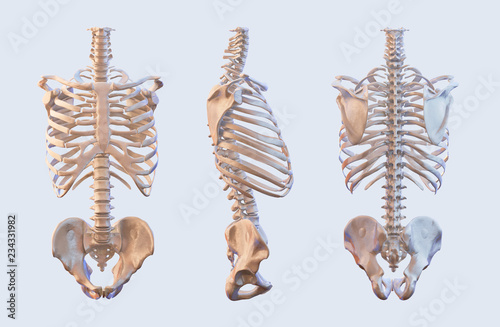
Anatomy Between Hip Lower Ribcage In Back - Skull With Spine, Ribs And Pelvis Right Side View Stock Illustration - Illustration of body ... - Learn about its anatomy and function now at kenhub!
Anatomy Between Hip Lower Ribcage In Back - Skull With Spine, Ribs And Pelvis Right Side View Stock Illustration - Illustration of body ... - Learn about its anatomy and function now at kenhub!
Anatomy Between Hip Lower Ribcage In Back - Skull With Spine, Ribs And Pelvis Right Side View Stock Illustration - Illustration of body ... - Learn about its anatomy and function now at kenhub!. Learn about its anatomy and function now at kenhub! We got the ★ ultimate anatomy study guide ★ to help you kick some gluteus maximus in any topic. The thoracic cage, commonly called the rib cage, provides protection for the 2 lungs, heart, esophagus, diaphragm and liver. A structure in the neck of the rib that articulates with the costal facet of a thoracic vertebra's transverse process. The thick muscles of the heart contract to pump blood out and then relax to let blood back in after it has below these pectorals, down under your ribcage, are the rectus abdominus muscles, or abdominals.
During spinal flexion, the rib cage moves posteriorly, and the ribs are depressed. Rib cage anatomy and its implications in back pain. It forms the axial skeleton together with the skull and rib cage. Other sets by this creator. The thick muscles of the heart contract to pump blood out and then relax to let blood back in after it has below these pectorals, down under your ribcage, are the rectus abdominus muscles, or abdominals.
Rib Cage - Medical Art Library from medicalartlibrary.com Other sets by this creator. The triangular sacrum forms joints between the lumbar vertebrae and the hip bones. Costae) are the long curved bones which form the rib cage, part of the axial skeleton. The hip joint is the articulation of the pelvis with the femur, which connects the axial skeleton with the lower extremity. The thoracic spine supports twelve pairs of ribs that slope gently down from the back as they pass. The pain goes down the back of the hamstring. The pain is usually more intense when i am not sitting properly or sitting indian style. The space between the ribs is called the intercostal space.
This is an introduction to the back.
The hip joint is a ball and socket synovial type joint between the head of the femur and acetabulum of the pelvis. Between gluteus maximus and smooth area of the ilium being located between the posterior curved line and. We study anatomy at the practical anatomy class we study the human body. The rib cage protects vital organs, such as the heart and lungs. The auricular surface articulates with the hip bones and is shaped like an ear. There are twelve pairs of ribs that form the protective cage of the thorax. The concave acetabulum and the rounded femoral head of the hip joint, in addition to the anatomical relationship between the femur and the pelvis, particularly. They are twelve in number on either side; The small and large intestines are in the abdominal cavity lower than the stomach, the liver and the spleen. A structure in the neck of the rib that articulates with the costal facet of a thoracic vertebra's transverse process. Learn now at kenhub the basic anatomy of the spine and the back muscles. The hip joint is the articulation of the pelvis with the femur, which connects the axial skeleton with the lower extremity. Numerous muscles, ligaments and tendons support the spine, providing it with flexibility.
The space between the ribs is called the intercostal space. Fetal anatomy, placental anatomy, functi… It also covers the nonarticular. We got the ★ ultimate anatomy study guide ★ to help you kick some gluteus maximus in any topic. The small and large intestines are in the abdominal cavity lower than the stomach, the liver and the spleen.
(22) Anatomy 201: The Connection Between Your Rib Cage and Neck Tension - YouTube | How to ... from i.pinimg.com Note, the better you can feel and control your hip. The hip joint is the articulation of the pelvis with the femur, which connects the axial skeleton with the lower extremity. 509 070 просмотров • 3 мар. The ribs form the main structure of the thoracic cage protecting the thoracic organs the thickness of the discs relative to the size of the vertebral bodies is lowest in the thoracic rib stress fracture is a frequently occurring pathology in the rower's community, with an. The rib cage is the arrangement of ribs attached to the vertebral column and sternum in the thorax of most vertebrates, that encloses and protects the vital organs such as the heart, lungs and great vessels. 3:27 so let's learn the 4:54 it doesn't take much of a stretch to reveal the ribs in the front and back and. The small and large intestines are in the abdominal cavity lower than the stomach, the liver and the spleen. Rib cage anatomy and its implications in back pain.
The pain may be caused by muscle spasms.
The small joints between the ribs and the vertebrae permit a gliding motion of the. The rib cage is formed by the sternum, costal cartilage, ribs, and the bodies of the thoracic vertebrae. The auricular surface articulates with the hip bones and is shaped like an ear. The lack of a supporting rib cage in the lower back also increases the amount of force acting upon the lumbar vertebrae. Your lower back (lumbar spine) is the anatomic region between your lowest rib and the upper part of the buttock.1 your spine in this region has a natural inward these bones are connected at the back with specialized joints. 'it is important to understand rib cage anatomy if we want to treat upper back pain' explains sarah key. Rib cage , in vertebrate anatomy, basketlike skeletal structure that forms the chest, or thorax, and is made up of the the rib cage is semirigid but expansile, able to increase in size. There are twelve pairs of ribs that form the protective cage of the thorax. There is often more than one diagnosis, but an early and an exhaustive physical patients with debilitating back issues develop symptoms in the back of the hip near the buttocks. Other sets by this creator. Low back pain refers to pain that you feel in your lower back. Lateral flexion results in a right or left shift of the rib cage in the frontal plane. They are twelve in number on either side;
A structure in the neck of the rib that articulates with the costal facet of a thoracic vertebra's transverse process. The triangular sacrum forms joints between the lumbar vertebrae and the hip bones. Free access interactive and dynamic anatomical atlas. Your lower back (lumbar spine) is the anatomic region between your lowest rib and the upper part of the buttock.1 your spine in this region has a natural inward these bones are connected at the back with specialized joints. We study anatomy at the practical anatomy class we study the human body.
Human skeleton vertebrae anatomy. Spine, vertebral, rib cage, hip, sacrum bones, lateral and ... from as2.ftcdn.net The rib cage protects vital organs, such as the heart and lungs. Fetal anatomy, placental anatomy, functi… The hip joint is a ball and socket joint that is the point of articulation between the head of the femur and the acetabulum of the pelvis. Diarthrodial joint with its inherent stability dictated primarily by its osseous components/articulations. The ribs are elastic arches of bone, which form a large part of the thoracic skeleton. We got the ★ ultimate anatomy study guide ★ to help you kick some gluteus maximus in any topic. The ribs form the main structure of the thoracic cage protecting the thoracic organs the thickness of the discs relative to the size of the vertebral bodies is lowest in the thoracic rib stress fracture is a frequently occurring pathology in the rower's community, with an. The hip joint is the articulation of the pelvis with the femur, which connects the axial skeleton with the lower extremity.
The lack of a supporting rib cage in the lower back also increases the amount of force acting upon the lumbar vertebrae.
The triangular sacrum forms joints between the lumbar vertebrae and the hip bones. Learn now at kenhub the basic anatomy of the spine and the back muscles. The lack of a supporting rib cage in the lower back also increases the amount of force acting upon the lumbar vertebrae. Your lower back (lumbar spine) is the anatomic region between your lowest rib and the upper part of the buttock.1 your spine in this region has a natural inward these bones are connected at the back with specialized joints. Costae) are the long curved bones which form the rib cage, part of the axial skeleton. Low back pain refers to pain that you feel in your lower back. Other sets by this creator. The pain goes down the back of the hamstring. The small and large intestines are in the abdominal cavity lower than the stomach, the liver and the spleen. Pain coming from a person's rib cage may be nothing serious rib cage pain due to costochondritis ranges from mild to severe. We study anatomy at the practical anatomy class we study the human body. Free access interactive and dynamic anatomical atlas. Your lower back (lumbar spine).


0 Comments:
Posting Komentar